Sunday, September 19, 2010

Sample for our assignment/learning topic help ! Epethilial tissue

EPETHILIAL TISSUE
Epethilial tissue covers the whole surface of the body.It covers body surfaces and lines hollow organs, body cavityies and ducts.It also forms glands
An epethilial tissue consist of cells arranged in continuos sheets, in either single or multiple layers.As cells are closely packed and are held tightly together by many cell junctions, there is little intercellular space between adjacent plasma memberanes.
Epethilial tissue may be divided inti two types :

  1. Covering and lining epethilium : It forms the outer covering of skin and some internal organs.It also forms the inner lining of blood vessels, ducts and body cavities, and the interior of respiratory tract, digestive tract, urinary tract and reproductive tract. Epithelial tissue that occurs on surfaces on the interior of the body is known as endothelium 
  2. Glandular epethilium : It makes up the secretin portion of the glands such as thyroid gland, adrenal gland and sweat gland.
The covering and lining epethilium tissue is further classified according to two characteristics like the arrangement of cells into layers and the shapes of the cells.

According to arrangement of cells into layers :

SIMPLE EPETHILIUM : Layer of cells that functions in diffussion, osmosis, filteration, secretion, absorption.It may be:

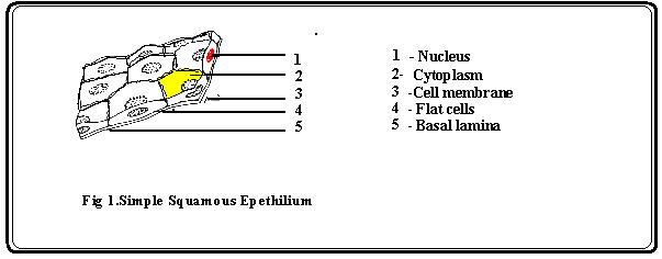
  1. Simple squamous (pavement) epithelium
Description : Single layer of flat cells, Centrally located nucleus.
Location : Lines heart, Blood vessels,Lymphatic vessels, Air sacs of lungs. Glomerular (Bowman's capsule) of kidney, Inner surface of tympanic membrane( Ear drum),
Functions : Filtration, diffussion ,Osmosis and Secretion into serous memberanes


      2.  Simple Cuboidal Epithelium

Description : Single layer of cube shaped cells, Centrally located nucleus.
Location : Covers the surface of ovary, Lines anterior surface of capsule of lens of eye, Lines kidney tubules and smaller ducts of many glands, Makes up secretion portion of thyroid gland and duct of pancreas.
Functions : Secretion and Absorption.


      3   Simple Columnar Epithelium
Description : One or more layers, Elongated and Column-shaped and Elongated nuclei  located near the base of the cells.Goblet cells (unicellular glands) are found between the columnar epithelial cells of the duodenum. They secrete mucus or slime, a lubricating substance which keeps the surface smooth. 
Location : Lining of the stomach and intestines, Specialised for sensory reception such as in the nose, ears and the taste buds of the tongue.
Functions : Secretion and Absorption.


     4. Ciliated Columnar Epithelium
Description : simple columnar epithelial cells, in addition, they posses fine hair-like outgrowths, cilia on their free surfaces.
Location : Air passages like the nose,Uterus and Fallopian tubes of females
Functions : Capable of rapid, rhythmic, wavelike beatings in a certain direction


STRAITED EPETHILIUM
  
       1.   Stratified squamous epithelium.
Description : Several layers of cells cuboidal to columnar shape in deep layers.
Location : Keratinized variety forms superficial layer of skin, nonkeratinized variety lines wet surface such as lining of mouth , eosophagus.
Function : Protection.


       2. Straitified cuboidal epethilium
Description : Two or more layers of cells in which the cells in the apical layer is in cube shaped.
Location : Ducts of adult sweat glands and esophageal glands and part of male urethera.
Function : Protection, Limited secretion and Absorption.


         3. Straitified columnar epethilium
Description : Several layers of irregularly shaped cells, only the apical layer has columnar cells.
Location : Lines part of Urethra, Large excretory ducts of some glands such as esophagus glands, small areas of anal mucous membrane, and par of the conjunctiva of the eye.
Function : Protection

     4. Transitional epethilium
Description : Appearance is variable (transitional), shape of cells in apical layer ranges from sqamous (when stretched ) to cuboidal (when relaxed).
Location : Lines urinary bladder and portions of ureters and urethra.
Function : Permts distension.


GLANDULAR EPETHILIUM

     1.Endocrine glands
Description : Secretory products (hormones) diffuse into blood after passing through interstitial fluid.
Location : Pituitary glan at base of brain, Pineal gland in brain, Thyroid and Parathyroid gland near larynx, Adrenal glands superior of kidneys.
Function : Produce hormone that regulate various body activities.
    2. Exocrine glands
Description : Secretory products released into ducts.
Location : Sweat, Oil and Earwax glands of the skin, Digestive glands such as salivary glands and pancreas.
Function : Produce substance such as Sweat , Oil earwax , Saliva and Digestive enzymes.


Monday, September 13, 2010

Chek out our Assignment help ! Assignment on Life cycle of viruses

LIFE CYCLE OF VIRUSES


INTRODUCTION TO VIRUSES

Viruses are simple, non-cellular entities consisting of one or more molocules of either DNA or RNA enclosed in a coat of protein.Some viruses are enveloped by capsid some are naked. They can reproduce only within living cells and are obligate intra-cellular parasites. Viruses are smaller than prokaryotic cells ranging in size from 0.02 to 0.03 micrometre. A fully assembled infectious virus is called a Virion.The main function of virion is to deliver its DNA or RNA genome into the host cell so that the genome can be expressed by the host cell.Each viral species has very limited host range.


TYPES OF VIRUSES BASED ON HOST

Prokaryotic viruses : Viruses that infect prokaryotes like bacteria.

Eukaryotic viruses : Viruses that infect Eukaryotes like animals and plants.


LIFE CYCLE OF PROKARYOTIC VIRUSES(BACTERIOPHAGE)-  The general life cycle of prokaryotic viruses are examplified by bacteriophage.For a virus to replicate it must induce a living host cell to synthesys all essential components needed to make more virions.These components must then be assembled into new virions and escape from the cell in order to infect other cells. There are to different viral life cycle Lytic and Lysogenic.


LYTIC LIFE CYCLE

Lytic or vegitative life cycle culminates in lysis or rupture of host cell and release numerous viral progeny. These are also called as virulent bacteriophages because they cause death of bacteria.

Lytic life cycle consists of following steps:

• Adsorption or docking with the host receptor protein.

• Entry or peneteration of the viral nucleic acid into the host cytoplasm.

• Biosynthesis of the viral components.

• Assembly (Maturation) of the viral components into complete viral units. After assembly viral proteins cause the lysis of host cell.

• Release of the completed virus from the host cell.

Example of Lytic phage :- T-even phages.


LYSOGENIC LIFE CYCLE

In the Lysogenic cycle the viral DNA is inserted into the host's DNA and it reproduces as the host's DNA replicates. A lysogenis virus can remain in this state for numerous replications of host cell DNA untill it excises itself from the host DNA and undergo a lytic life life cycle.When the genetic material of these phages is inserted into the DNa of host cells it is said to be in the prophage state. A cell that contains a prophage is known as lysogen.These are also called as temperate phages.

Example: lambda phage


SHOWING PROKARYOTIC VIRUSE LIFE CYCLE (LYTIC AND LYSOGENIC)


LIFE CYCLE OF EUKARYOTIC VIRUSES

ENVELOPED VIRUSES

• Adsorption begins with the docking proteins of the virus locking into specific receptor sites on the surface of the target host cells.

• In the case of some enveloped eukaryotic viruses the viral membrane fuses with the cell membrane and in effect opens up so as to allow the virus to enter the cell cytoplasm. Once in the cytoplasm the protein capsid is removed and the viral genome is freed into the cytoplasm. Depending on the nature of the virus the viral genome may stay in the cytoplasm or it may enter the nucleus before beginning the next stage of the life cycle.

• In another case the cell engulfs the entire enveloped eukaryotic virus, taking it into the cytoplasm,called as endocytosis. Once inside the cytoplasm the enveloped eukaryotic virus is said to be in a vesicle. Subsequently, the two lipid membranes of the vesicle fuse, releasing the capsid covered virus. The capsid is then removed and the viral genome is freed for the next stage of viral replication.

• The actual processes from biosynthesis to maturation and release varies depending on the virus type. However, at the core, the viral genomic material uses cellular metabolism for its own selfish purposes. Finally the mature virus or virions. are released into the environment to begin the cycle again.


SHOWING EUKARYOTIC VIRUS (RETROVIRUS)LIFE CYCLE


NAKED VIRUSE

The naked virus replication is less elaborate, but follows the same course. After binding to the host receptors the naked viral capsid penetrates into the host cytoplasm, the genome is released and the steps of viral replication ensue. Since the final release of enveloped eukaryotic viruses involves taking a portion of the cell membrane the process is called budding. For example, the HIV virus buds out of the cell. Whether release involves lysis or budding, a cell eventually dies due to the viral infection and destruction of its metabolism


Examples of eukaryotic viruses are :

Animal viruses - adenovirus, retrovirus

Plant viruses - tobacco mosaic virus

Insect viruses - baculovirus

SUMMARY

Viruses replicate inside the host using host machinary. Different viruses replicate in specific host by differenet mechanisms.General life cycle of viruses include genome entry to host and then replicate in host , assembly of new and then release.